Service.
We offer a complete set of high-quality diagnostic services.
Our equipment is ultra-modern
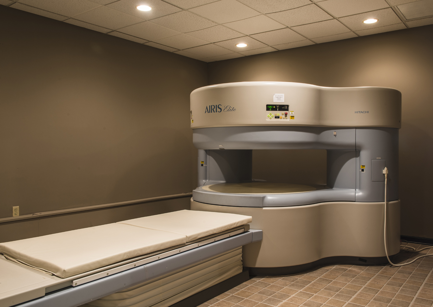
MRI SCAN

What is MRI SCAN?
Magnetic resonance imaging (MRI) is a technique that uses a magnetic field and radio waves to create detailed images of the organs and tissues within your body.
When is MRI required?
MRI scan can help to diagnose the following:
- most ailments of the brain, including tumours and dementias
- sports injuries
- musculoskeletal problems
- most spinal conditions/injuries
- vascular abnormalities
- female pelvic problems
- prostate problems
- some gastrointestinal tract conditions
- certain ear, nose and throat (ENT) conditions
- soft tissue and bone pathology/conditions
Are there any risks associated with MRI?
Because MRI uses powerful magnets, the presence of metal in your body may be a safety hazard or affect a portion of the MRI image. Before having an MRI, tell the technologist if you have any metal or electronic devices in your body, such as:
- Metallic joint prostheses
- Artificial heart valves
- An implantable heart defibrillator
- A pacemaker
- Metal clips
- Cochlear implants
- A bullet, shrapnel or any other type of metal fragment
CT SCAN
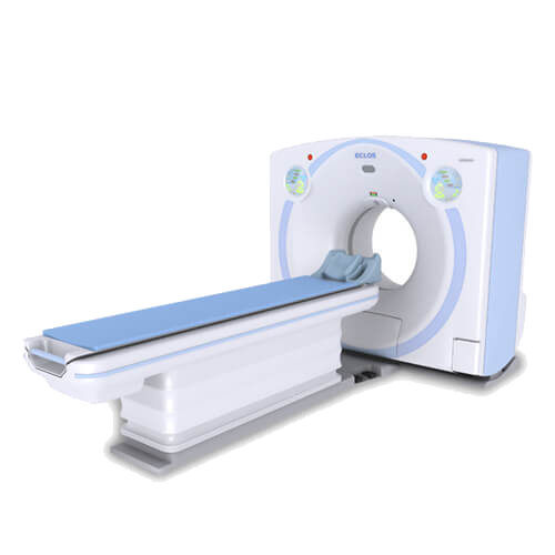
What is CT scan?
A computerized tomography (CT) scan combines a series of X-ray images taken from different angles and uses computer processing to create cross-sectional images, or slices, of the bones, blood vessels and soft tissues inside your body. CT scan images provide more detailed information than plain X-rays do.
A CT scan has many uses, but is particularly well-suited to quickly examine people who may have internal injuries from car accidents or other types of trauma. A CT scan can be used to visualize nearly all parts of the body and is used to diagnose disease or injury as well as to plan medical, surgical or radiation treatment.
Why is it done?
Your doctor may recommend a CT scan to help:
- Diagnose muscle and bone disorders, such as bone tumors and fractures
- Pinpoint the location of a tumor, infection or blood clot
- Guide procedures such as surgery, biopsy and radiation therapy
- Detect and monitor diseases and conditions such as cancer, heart disease, lung nodules and liver masses
- Monitor the effectiveness of certain treatments, such as cancer treatment
- Detect internal injuries and internal bleeding
Does it require any special preparation?
Depending on which part of your body is being scanned, you may be asked to:
- Take off some or all of your clothing and wear a hospital gown
- Remove metal objects, such as a belt, jewelry, dentures and eyeglasses, which might interfere with image results
- Refrain from eating or drinking for a few hours before your scan
Contrast material
A special dye called a contrast material is needed for some CT scans, to help highlight the areas of your body being examined. The contrast material blocks X-rays and appears white on images, which can help emphasize blood vessels, intestines or other structures.
Contrast material might be given to you:
- By mouth. If your esophagus or stomach is being scanned, you may need to swallow a liquid that contains contrast material. This drink may taste unpleasant.
- By injection. Contrast agents can be injected through a vein in your arm to help your gallbladder, urinary tract, liver or blood vessels stand out on the images. You may experience a feeling of warmth during the injection or a metallic taste in your mouth.
MAMMO-
GRAPHY
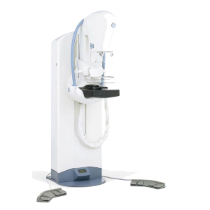
What is mammography or mammogram?
A mammography, or mammogram, is an X-ray of the breast. It’s a screening tool used to detect and diagnose breast cancer. Together with regular clinical exams and monthly breast self-examinations, mammograms are a key element in the early diagnosis of breast cancer.
According to the National Cancer Institute, breast cancer is the second most common cancer for women in the United States, after skin cancer. There are about 2,300 new cases of breast cancer in men each year, and about 230,000 new cases in women each year.
Some experts recommend that women who are 40 years old and older should have mammograms every one to two years. The American Cancer Society recommends regular screening beginning at age 45. If you have a personal or family history of breast cancer, your doctor may recommend that you start screenings earlier, have them more often, or use additional diagnostic tools.
If your doctor orders a mammogram as a routine test to check for any cancer or changes, it’s known as a screening mammogram. In this type of test, your doctor will take several X-rays of each breast.
If you have a lump or any other symptom of breast cancer, your doctor will order a diagnostic mammogram. If you have breast implants you will probably need a diagnostic mammogram. Diagnostic mammograms are more extensive than screening mammograms. They typically require more X-rays to get views of the breast from multiple positions. Your radiologist may also magnify certain areas of concern.
How Do I Prepare for a mammography or mammogram?
You will need to follow certain guidelines on the day of your mammogram. You can’t wear deodorants, body powders, or perfumes. Also, you shouldn’t apply any ointments or creams to your breasts or underarms. These substances can distort the images or look like calcifications, or calcium deposits, so it’s important to avoid them.
Be sure to tell your radiologist before the exam if you’re pregnant or breastfeeding. In general, you won’t be able to receive a screening mammogram at this time, but if necessary, your doctor can order other screening methods, such as an ultrasound.
What Happens During a Mammography?
After undressing from the waist up and taking off any necklaces, a technician will give you a smock or gown that ties in the front. Depending on the testing facility, you may either stand or sit during your mammogram.
Each breasts fits onto a flat X-ray plate. A compressor will then push the breast down to flatten the tissue. This provides a clearer picture of the breast. You might have to hold your breath for each picture. You may feel a small amount of pressure or discomfort, but it’s usually brief.
During the process, your doctor will review the images as they are made. They may order additional images that show different views if something is unclear or needs further attention. This happens quite frequently and shouldn’t be a cause for upset or panic.
Digital mammograms are sometimes used if they are available. These are especially helpful for women younger than 50 years old, who typically have denser breasts than older women.
A digital mammogram transforms the X-ray into an electronic picture of the breast that saves onto a computer. Images are immediately visible, so your radiologist doesn’t have to wait for the images. The computer can also help your doctor see images that might not have been very visible on a regular mammogram.
ULTRA-
SOUND
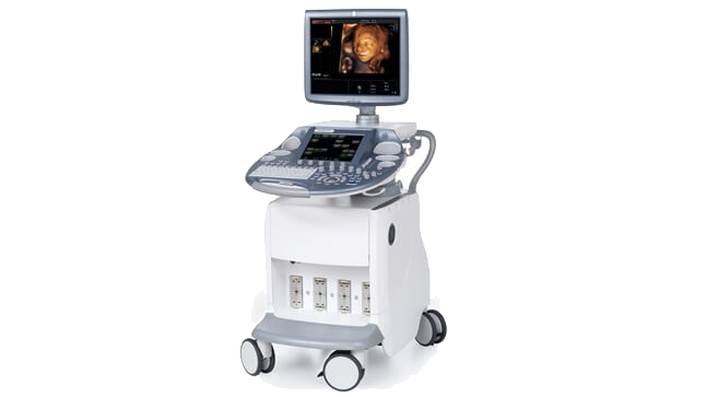
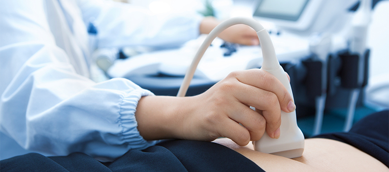
What is an ultrasound?
An ultrasound scan is a medical test that uses high-frequency sound waves to capture live images from the inside of your body. It’s also known as sonography.
The technology is similar to that used by sonar and radar, which help the military detect planes and ships. An ultrasound allows your doctor to see problems with organs, vessels, and tissues without needing to make an incision.
Unlike other imaging techniques, ultrasound uses no radiation. For this reason, it’s the preferred method for viewing a developing fetus during pregnancy.
How is an ultrasound performed?
Before the exam, you will change into a hospital gown. You will most likely be lying down on a table with a section of your body exposed for the test.
An ultrasound technician, called a sonographer, will apply a special lubricating jelly to your skin. This prevents friction so they can rub the ultrasound transducer on your skin. The transducer has a similar appearance to a microphone. The jelly also helps transmit the sound waves.
The transducer sends high-frequency sound waves through your body. The waves echo as they hit a dense object, such as an organ or bone. Those echoes are then reflected back into a computer. The sound waves are at too high of a pitch for the human ear to hear. They form a picture that can be interpreted by the doctor.
Depending on the area being examined, you may need to change positions so the technician can have better access.
After the procedure, the gel will be cleaned off of your skin. The whole procedure typically lasts less than 30 minutes, depending on the area being examined. You will be free to go about your normal activities after the procedure has finished.
E.C.G
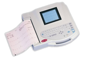
What is Electrocardiogram ( ECG) ?
An electrocardiogram records the electrical signals in your heart. It’s a common test used to detect heart problems and monitor the heart’s status in many situations. Electrocardiograms — also called ECGs or EKGs — are often done in a doctor’s office, a clinic or a hospital room. And they’ve become standard equipment in operating rooms and ambulances.
An ECG is a noninvasive, painless test with quick results. During an ECG, sensors (electrodes) that can detect the electrical activity of your heart are attached to your chest and sometimes your limbs. These sensors are usually left on for just a few minutes.
Why is an electrocardiogram done?
An electrocardiogram is a painless, noninvasive way to help diagnose many common heart problems in people of all ages. Your doctor may use an electrocardiogram to detect:
- Irregularities in your heart rhythm (arrhythmias)
- If blocked or narrowed arteries in your heart (coronary artery disease) are causing chest pain or a heart attack
- Structural problems with your heart’s chambers
- A previous heart attack
- How well certain ongoing heart disease treatments, such as a pacemaker, are working
You may need a heart rhythm test if you experience any of the following signs and symptoms:
- Heart palpitations
- Rapid pulse
- Chest pain
- Shortness of breath
- Dizziness, lightheadedness or confusion
- Weakness, fatigue or a decline in ability to exercise
Are there any risks associated with electrocardiogram?
An electrocardiogram is a safe procedure. You’ll have no risk of getting an electrical shock during the test because the electrodes placed on your body don’t emit electricity. They only record the electrical activity of your heart.
You may have minor discomfort, similar to removing a bandage, when the electrodes are removed. Rarely, a reaction to the electrode adhesive may cause redness or swelling where the patches were placed.
X-RAY
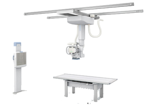
What is X-RAY?
An X-ray is a quick, painless test that produces images of the structures inside your body — particularly your bones.
X-ray beams pass through your body, and they are absorbed in different amounts depending on the density of the material they pass through. Dense materials, such as bone and metal, show up as white on X-rays. The air in your lungs shows up as black. Fat and muscle appear as shades of gray.
For some types of X-ray tests, a contrast medium — such as iodine or barium — is introduced into your body to provide greater detail on the images.
Why is it done?
X-ray technology is used to examine many parts of the body.
Bones and teeth
- Fractures and infections. In most cases, fractures and infections in bones and teeth show up clearly on X-rays.
- Arthritis. X-rays of your joints can reveal evidence of arthritis. X-rays taken over the years can help your doctor determine if your arthritis is worsening.
- Dental decay. Dentists use X-rays to check for cavities in your teeth.
- Osteoporosis. Special types of X-ray tests can measure your bone density.
- Bone cancer. X-rays can reveal bone tumors.
Chest
- Lung infections or conditions. Evidence of pneumonia, tuberculosis or lung cancer can show up on chest X-rays.
- Breast cancer. Mammography is a special type of X-ray test used to examine breast tissue.
- Enlarged heart. This sign of congestive heart failure shows up clearly on X-rays.
- Blocked blood vessels. Injecting a contrast material that contains iodine can help highlight sections of your circulatory system to make them visible on X-rays.
Abdomen
- Digestive tract problems. Barium, a contrast medium delivered in a drink or an enema, can help reveal problems in your digestive system.
- Swallowed items. If your child has swallowed something like a key or a coin, an X-ray can show the location of that object.
Are there any risk involved?
Some people worry that X-rays aren’t safe because radiation exposure can cause cell mutations that may lead to cancer. The amount of radiation you’re exposed to during an X-ray depends on the tissue or organ being examined. Sensitivity to the radiation depends on your age, with children being more sensitive than adults.
Generally, however, radiation exposure from an X-ray is low, and the benefit from these tests far outweigh the risks.
However, if you’re pregnant or suspect that you may be pregnant, tell your doctor before having an X-ray. Though the risk of most diagnostic X-rays to an unborn baby is small, your doctor may consider another imaging test, such as ultrasound.
Still need help? Call us
For any questions, please write us at info@dclgh.com
or call us on +233 0302211930